SAN DIEGO, CA.- Gastroesophageal reflux disease, more commonly known as GERD, impacts around 20 percent of U.S. citizens, according to the National Institutes of Health. If left untreated, GERD can lead to serious medical issues and sometimes esophageal cancer. Thanks to supercomputers, advances in imaging the swallowing process of GERD patients have been modelled on Comet at the San Diego Supercomputer Center (SDSC) at
UC San Diego and Bridges-2 at the Pittsburgh Supercomputing Center (PSC).
Northwestern University researchers from the McCormick School of Engineering and the Feinberg School of Medicine teamed up and recently published these novel models in Biomechanics and Modeling in Mechanobiology. Their work resulted in a new computational modeling system, called FluoroMech, that could help identify physio-markers for early and accurate diagnosis of esophageal pathologies.
“While nearly 60 percent of the adult population experiences some form of GERD each year, there are 500,000 new cases of esophageal cancer diagnosed each year worldwide, with a projected 850,000 new cases/year by 2030,” said Neelesh Patankar, a professor of mechanical engineering at Northwestern and the study’s senior author. “In the U.S., there are nearly 17,000 diagnoses per year, accounting for about one percent of cancer diagnoses, but less than 20 percent of these patients survive at least five years and the primary curative treatment is esophagectomy.”
Patankar said that staggering statistics such as these for esophageal disorders in general inspired the team of engineering and medical researchers to create an interdisciplinary study, which resulted in FluoroMech, that complements common non-invasive medical imaging techniques to quantitatively assess the mechanical health of the esophagus.
Why it’s important
Mechanical properties, such as elasticity, of the esophageal wall and its relaxation during swallowing have been shown to play a critical role in the functioning of the esophagus; thus, these qualities have been considered as indicators, or physio-markers, that explain the health of the organ. Because diagnostic techniques that determine these physical quantities are lacking, the Northwestern research team developed the FluoroMech computational technique to help clinicians have more thorough and accurate information about each patient’s esophagus.
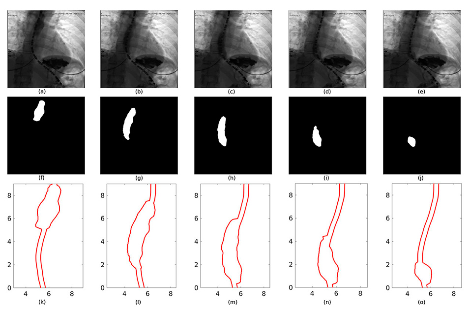
Specifically, FluoroMech was designed to predict mechanical properties of soft tubular organs such as the esophagus by analyzing medical images and video from fluoroscopy (real-time X-ray imaging) of the esophagus during a swallowing process. The new technique has also been developed to enable the clinician to quantify the relaxation of the esophageal muscles during the passage of food.
“An understanding of mechanisms underlying human pathophysiology requires knowledge of both biochemical and biomechanical function of organs—yet the use of biomechanics has not advanced on par with biochemistry or even imaging techniques like MRIs and X-rays,” said Patankar. “This is particularly true in the case of esophageal disorders, and we aim to shift the existing paradigm of studying disease pathogenesis of organs toward a biomechanics-based approach that harnesses information about how mechanical properties of organs alter physiology.”
How supercomputers helped
“In our biomechanics problem, the entire system had almost five million unknowns to solve at each time step and there are large numbers of time steps that needed to be solved—simulations of this magnitude required advanced supercomputers to obtain results in a reasonable time, which is often five to seven days for each model,” explained Sourav Halder, a Northwestern doctorate student and the study’s lead author. “Without the availability of these computational resources, it would not have been possible to simulate such systems.”
These recent supercomputer-enabled models furnished the team with the tools to accomplish its lofty goal of creating FluoroMech. Prior to the development of its technique, it was not possible to quantify the mechanical health of the esophagus, and perhaps most importantly allow for patient-specific predictive modelling. Halder said this was previously impossible due to the lack of automated image segmentation based on machine learning techniques and complex physics-based calculations of esophageal function. With the help of supercomputers that are part of the National Science Foundation Extreme Science and Engineering Discovery Environment (XSEDE) as well as the education and training team at SDSC, Halder and colleagues were able to build out their FluoroMech technique.
Halder applauded the SDSC support team for its help in setting up proprietary software to enable efficient prototyping of simulation models and the ability to generate massive amounts of data for machine learning models. He said that the SDSC team also offered educational seminars on GPU Computing, HPC in Data Science and high-level programming frameworks like OpenACC. “We are extremely thankful to the SDSC education and training team for regularly conducting these workshops that are extremely accessible to everyone in attendance irrespective of their computational background,” he said.
What’s next?
“While FluoroMech uses fluoroscopy data to predict esophageal wall properties as well as the functioning of the esophagus in terms of estimating active relaxation of the muscle walls, we have recently extended our work with another diagnostic device called EndoFLIP (Endolumenal Functional Lumen Imaging Probe) to predict mechanics-based physio-markers such as the esophageal wall contraction strength, active relaxation and wall elastic properties,” said Patankar.
Using EndoFLIP data from a large cohort of subjects and the predictions from the FluoroMech model, the team has started to develop a virtual disease landscape (VDL). The VDL is a parameter space where subjects of different esophageal disorders get clustered into different regions. The locations of the clusters, relative to each other, are designed to represent similarities and differences between the modes of bolus transport through the esophagus. The prototypes of the VDL concept has already shown how it can provide fundamental understanding of the underlying physics of various esophageal disorders.
“Thanks to XSEDE allocations, we have already been able to develop improved models for gastric peristalsis, illustrate muscular activity in the stomach and simulate acid reflux,” said Halder. “Newer clusters like Expanse at SDSC were built with double the amount of RAM on each node and nearly five times the number of cores compared to Comet, so this leap in computational power lets us plan for advanced models without seeing a substantial increase in time required for computation.”
Support for this research was provided by grants from Public Health Service (R01-DK079902 and P01-DK117824) and the National Science Foundation (NSF) (OAC 1450374 and OAC 1931372). Computational resources were provided by Northwestern University’s Quest High Performance Computing Cluster and the Extreme Science and Engineering Discovery Environment (XSEDE) through allocation TG-ASC170023, which is supported by NSF (ACI-1548562). It also used SDSC’s Comet, which is supported by NSF (ACI-1548562) and the Bridges-2 system at PSC, which is supported by NSF (ACI-1928147).